
Physiologic Basic of Human Eye
The optical model of the human eye describes the basic components and their functions in focusing light onto the retina. Figure 1 is a simplified explanation of the optical model of the human eye:
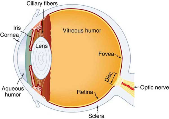
Figure 1: General human eye structure
- Cornea: The cornea is the transparent front surface of the eye. It acts as a fixed lens and is primarily responsible for bending (refracting) incoming light.
- Iris and Pupil: The iris is the colored part of the eye that surrounds the pupil, which is the adjustable opening in the center of the iris. The iris controls the size of the pupil, regulating the amount of light that enters the eye.
- Lens: Behind the pupil, flexible, transparent lens. The lens can change shape to adjust focusing power, allowing for accommodation and the ability to focus on objects at different distances.
- Aqueous Humor: The space between the cornea and the lens is filled with a clear fluid called aqueous humor. It helps maintain the shape of the eye and provides nutrients to the cornea and lens.
- Vitreous Humor: The main chamber of the eye, behind the lens, is filled with a gel-like substance called the vitreous humor. The vitreous humor helps maintain the shape of the eye and supports the retina.
- Retina: The innermost layer at the back of the eye. It contains specialized cells called photoreceptors (rods and cones) that detect light and convert it into electrical signals. These signals are then sent to the brain via the optic nerve for further processing and interpretation.
Optical Model of Human Eye in Zemax OpticStudio:
The Eye Model in Ansys Zemax OpticStudio is a computational representation of the human eye that enables the simulation of light propagation through the eye's optical components, such as the cornea, lens, humors and retina. It provides a means to study and analyze various aspects of vision, including image formation, aberrations, and visual performance. Here presents an eye model illustration in Zemax Sequential Mode, with parallel beam in multiple fields (0°, 15°, 30°) as incidences. The weights of these fields are 1, 0.3, 0.1 in image analysis. Spectrum covers wavelength in 470-650 nm in 4 steps, with the middle step 555 nm as the maximum weight of 1.
Components of eye depicted above are edited in Lens Data Editor of sequential model to reflect the eye structure. The key components are marked in Figure 2 below. Optical properties of bio-organ are depicted by Conrady formula,
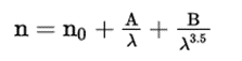
which can be found in Zemax user manual. Detailed numbers are listed in Table 1.
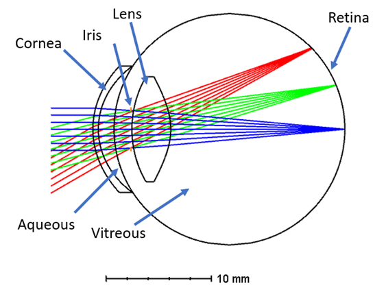
Figure 2: Human eyeball model in Ansys Zemax sequential mode
Table 1: Refractive index and dispersion coefficients of eye organs
|
|
n0 |
A |
B |
|
Aqueous |
1.32 |
8.47E-3 |
2.31E-2 |
|
Lens |
1.40 |
9.39E-3 |
3.93E-4 |
|
Cornea |
1.63 |
6.67E-3 |
3.87E-4 |
|
Vitreous |
1.32 |
6.73E-3 |
3.34E-4 |
Here we take a scene with multiple color (Figure 3 left) as an incident beam to the eyeball. In the retina as detector, 88.7% of light is received and form the image below at Figure 3 right. The spot diagram is shown in Figure 4. Geometric modulation transfer function (MTF) up to 20 lp/mm is shown in Figure 5.

Figure 3: Left, original scene as a remote object; Right, image at retina.

Figure 4: Standard spot diagram

Figure 5: Geometric modulation transfer function
Using the Zemax Eye Model, optical designers can evaluate the performance of vision correction techniques like eyeglasses, intraocular lenses or AR/VR products. It allows to simulate the effect of different refractive errors, such as myopia (nearsightedness), hyperopia (farsightedness), astigmatism, and presbyopia (age-related near vision decline). By manipulating the model parameters, researchers can investigate the impact of these conditions on visual acuity, image quality, and other optical properties.
