
Laser Speckle Contrast Imaging (LSCI) is a non-invasive imaging technique used primarily in biomedical research and clinical applications to visualize blood flow in tissues. It relies on the phenomenon of speckle contrast, which is the variation in the intensity of laser light scattered by moving red blood cells within biological tissues. By analyzing the contrast of the speckle pattern, LSCI can provide information about blood flow velocity and perfusion.
Illumination is a critical aspect of LSCI because it determines how laser light interacts with the biological tissue, creating the speckle pattern used to analyze blood flow. Proper illumination is essential for obtaining accurate and meaningful LSCI results.
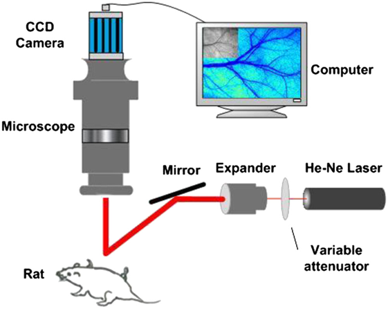
Fig.1 Laser speckle imaging configuration
There are some key considerations for the illumination setup in Laser Speckle Contrast Imaging. The first is wavelength. Typically, red or near-infrared wavelengths are used (e.g., 785 nm or 633 nm) because they can penetrate biological tissues with minimal absorption and scattering. Power is also considerable. The laser power should be adjustable to control the intensity of illumination on the tissue. The power level should be within safe limits to avoid tissue damage. Beam shape and size need to be controlled in the illumination as well. Use optical components like lenses, beam expanders, or diffusers to control the shape and size of the laser beam. The beam should be adjusted to cover the region of interest on the tissue and provide uniform illumination. The next is spatial coherence. Laser light is coherent, which means it can create sharp speckle patterns. In LSCI, it's often desirable to reduce the spatial coherence of the laser beam to obtain more informative speckle patterns. This can be achieved by introducing some degree of scattering, such as using ground glass or a diffuser in the optical path. One additional is illumination angle. The angle at which the laser beam illuminates the tissue can affect the speckle pattern's characteristics. Typically, an oblique angle is used to maximize the contrast in the speckle pattern.
Fig.2 illustrates the illumination part of LSCI. The laser source is modeled as a “Source Diode” type object in Zemax Non-sequential mode. The X- and Y-divergence angle are both 6 degree. Diffuser is modeled with a cylinder volume in a diameter of 1-inch, and thickness of 2 mm. Both front and rear surfaces are set as Lambertian scatter, with scattering proportion of 0.6. The distance between the diffuser and the illumination plane is 95 mm.
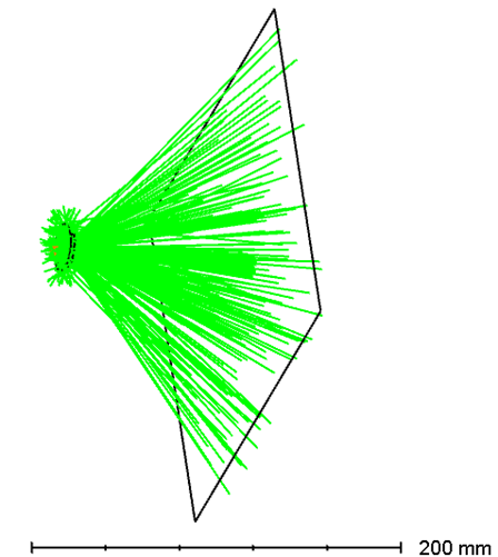
Fig.2 Illumination setup of LSCI
LSCI typically uses a coherent laser source, such as a diode laser, with a specific wavelength (e.g., 785 nm or 633 nm) that can penetrate biological tissues. The laser light is directed onto the tissue of interest using appropriate optics, which may include lenses, beam expanders, and diffusers to achieve the desired illumination area and intensity.
The size of the interested illumination area is 100 x 100 mm2. Fig. 3 shows the incoherence irradiance over the illumination area. Fig. 4 shows the illumination map at the specimen plane. The total received power is 0.33 Watt when the source input is set as 1 Watt. The total flux is 56 lumens. This typical structure can be used in LSCI system and related fluorescent illumination setups.
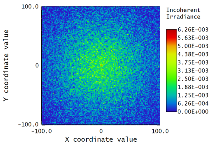
Fig.3 Irradiance map on the LSCI specimen plane
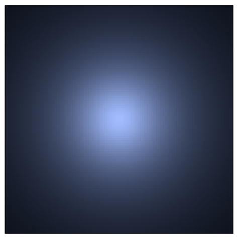
Fig.4 Illumination map of the LSCI specimen plane
